
Key Takeaways: From Dr. Prince Uchadiya’s Clinical Experience in Indore
- The most dangerous thing about a stress fracture is that it often looks like nothing on a plain X-ray. In my clinic in Nipania, Indore, I see patients who have been told their X-ray is normal and sent home, only to arrive weeks later with a complete fracture that could have been prevented. X-ray misses stress fractures in the early stages. MRI does not.
- Stress fractures are not all equal. There is a critical clinical distinction between low-risk fractures, which heal reliably with rest and activity modification, and high-risk fractures of the femoral neck, anterior tibial cortex, navicular, and fifth metatarsal, which can progress to complete fracture, nonunion, or avascular necrosis if managed incorrectly.
- Tibia is the most commonly fractured bone, accounting for up to 49% of all stress fractures in athletes, followed by tarsal bones at 25% and metatarsals at 9%. PMC Stress Fracture Review
- In Indore’s growing running and fitness community, I see stress fractures predominantly in runners who have increased their mileage too quickly before the Indore Marathon, in young cricketers and kabaddi players during pre-season intensification, and in female athletes with poor nutrition and low Vitamin D, which is nearly universal in central India.
- Stress fractures represent 15 to 20% of all musculoskeletal injuries in runners and up to 22% in female track-and-field athletes. PMC
- The average time to diagnosis in published studies is 13.4 weeks, meaning most patients have been in pain for over three months before the correct diagnosis is made. PubMed 320-Athlete Study
- The treatment decision is location-specific. Getting the location right determines whether the patient needs rest alone, non-weight-bearing immobilisation, or surgical fixation. Treating all stress fractures the same way is the most common reason they fail to heal.
- Vitamin D deficiency is a contributing factor I address in every stress fracture patient, because approximately 80% of urban Indians are deficient and it directly impairs bone remodelling capacity, extending healing time and raising re-fracture risk.
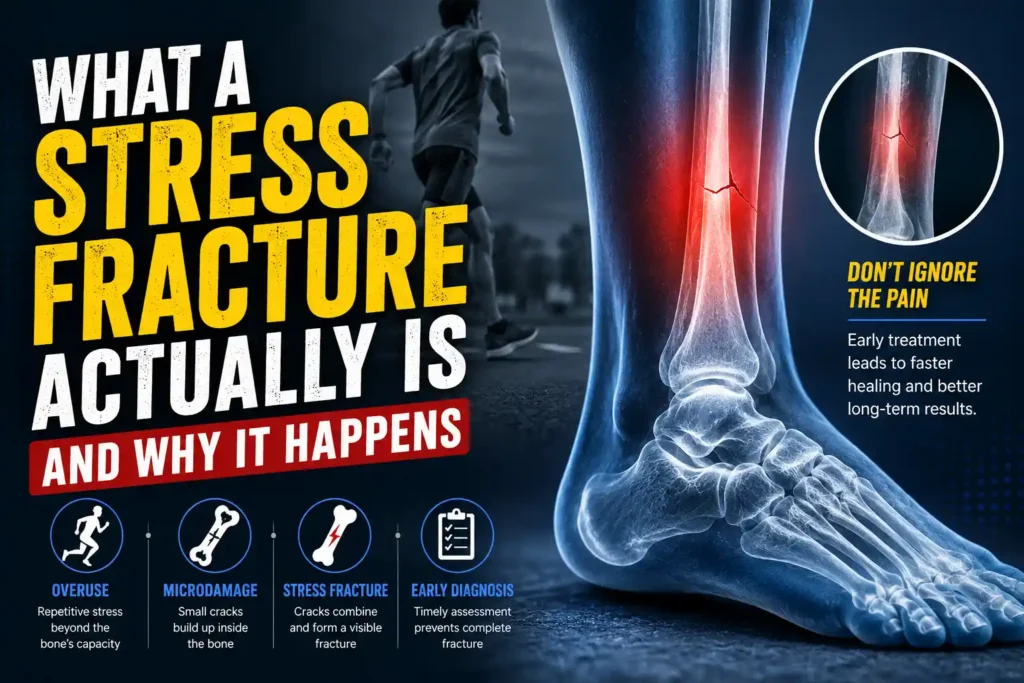
What a Stress Fracture Actually Is and Why It Happens
Bone is a dynamic structure. It is constantly being broken down by osteoclast cells and rebuilt by osteoblast cells in a process called remodelling. Under normal loading, this cycle maintains bone integrity. When repetitive mechanical load is applied faster than the bone can remodel and repair, microdamage accumulates. If the loading does not stop, those microcracks coalesce into a visible fracture line. This is a stress fracture.
Two distinct mechanisms produce stress fractures, and understanding the difference matters for treatment. The first is a fatigue fracture: normal bone subjected to abnormal, repetitive loading. This is the classic runner’s stress fracture, the soldier’s march fracture, the athlete who trained too hard too fast. The bone itself is structurally normal but the cumulative load has outpaced its repair capacity. The second is an insufficiency fracture: abnormal bone subjected to normal, everyday loading. This occurs in patients with osteoporosis, osteomalacia, or significant Vitamin D deficiency where the bone’s intrinsic strength is compromised. A patient with severe osteoporosis can develop a sacral insufficiency fracture simply from walking.
Both types result in the same pathology at the microscopic level but require different systemic management alongside the local fracture treatment. Identifying which mechanism is at work is part of the first consultation at Dr. Prince Uchadiya Orthopaedic And Joint Care Clinic in Nipania, Indore.
Who Gets Stress Fractures: The Indore Patient Profile
Stress fractures are not exclusively a problem of elite athletes. In practice in Indore, they appear across a wide range of patient types, and the profile has shifted significantly as recreational fitness culture has grown across the city.
Young runners preparing for the Indore Marathon who increase their weekly mileage by more than 10% per week represent the most common presentation. The tibia and metatarsals are the bones that fail first under this kind of rapid load increase. Young cricketers, particularly fast bowlers, develop lumbar pars stress fractures from the repetitive hyperextension loading of bowling action. Kabaddi players and court sports athletes develop tibial and metatarsal stress fractures from the explosive cutting and landing forces of their sport. Classical dancers develop metatarsal and sesamoid stress fractures from sustained weight-bearing through the forefoot. Military recruits and NDA aspirants in Indore who begin intensive physical training rapidly are another consistent group.
Female athletes carry a specific additional risk. NCBI StatPearls confirms that female athletes have a higher incidence of stress fractures than males in comparable training programmes. The female athlete triad, consisting of low energy availability, menstrual dysfunction, and low bone density, is a recognised contributing framework. A young female athlete who has reduced her food intake significantly while increasing training volume has essentially created the perfect conditions for a stress fracture.
Across all groups in Indore, widespread Vitamin D deficiency is a consistent background factor. With approximately 80% of urban Indians deficient in Vitamin D, bone remodelling capacity is compromised even in otherwise healthy patients. This extends healing time, raises re-fracture risk, and in some cases is itself the primary reason a patient has developed a stress fracture from training loads that most athletes manage without injury.
Symptoms That Should Trigger Immediate Assessment
The symptom pattern of a stress fracture is specific enough to be clinically recognisable before imaging confirms it. Pain that develops gradually over weeks rather than suddenly, that is directly related to a specific activity and worse toward the end of a training session, that improves with rest but returns immediately when training resumes, and that is precisely localised to one specific point on a bone rather than diffusely across a joint or muscle is the classic presentation.
Point tenderness on direct pressure over the fracture site is the most reliable clinical sign. In the tibia, pressing firmly along the bone shaft reproduces the pain at exactly the fracture location. In the metatarsals, pressing directly on the shaft of the affected bone reproduces it. The hop test, where the patient is asked to hop on the affected leg, is a useful provocation test that loads the bone axially and typically reproduces pain in tibial stress fractures within a few hops.
Night pain is a warning sign that warrants urgent imaging. Most stress fractures are silent at rest in the early stages. A fracture that is beginning to hurt at rest or at night has progressed beyond the early stage and may be approaching complete cortical disruption.
Swelling, warmth, and visible bruising overlying the fracture site can occur in more advanced cases, particularly in superficial bones like the tibia and metatarsals. Their absence does not exclude the diagnosis.
The Critical Distinction: High-Risk Versus Low-Risk Stress Fractures
This classification determines the entire treatment approach and is the most important clinical decision in stress fracture management. Getting it wrong, specifically treating a high-risk fracture as if it were low-risk, can result in complete fracture, nonunion, avascular necrosis, and a career-ending injury in an athlete or permanent disability in an older patient.
Low-Risk Stress Fractures
Low-risk stress fractures occur at sites where the bone is loaded in compression, has good blood supply, and has a reliable history of healing with conservative management. They include the posteromedial tibial diaphysis, metatarsal shafts (second and third), distal fibula, compression-side femoral neck, femoral shaft, and calcaneus. These fractures respond reliably to relative rest, activity modification, and gradual return to loading as symptoms resolve. Healing typically takes 6 to 12 weeks. The athlete does not need to stop all activity, just the specific loading pattern that caused the fracture.
High-Risk Stress Fractures
High-risk stress fractures occur at sites where the bone is loaded in tension, where blood supply is limited, or where the geometry of the fracture makes complete displacement or nonunion likely if loading continues. NCBI StatPearls identifies the high-risk locations as follows:
- Tension-side femoral neck: The most serious stress fracture in athletes. If this fractures completely, it can displace and cause avascular necrosis of the femoral head, a potentially career-ending and life-altering complication. Surgical fixation is the standard of care for tension-sided femoral neck stress fractures.
- Anterior tibial cortex: The “dreaded black line.” This site is under tensile loading and has notoriously poor healing due to the watershed blood supply. Conservative management has very low success rates. Surgical intramedullary nailing is frequently required.
- Tarsal navicular: Located in a watershed zone of poor blood supply and under significant compression loading as the keystone of the medial longitudinal arch. A missed or undertreated navicular stress fracture can progress to nonunion and chronic midfoot pain. Non-weight-bearing cast immobilisation for 6 to 8 weeks is the minimum conservative treatment. Displaced or nonuniting fractures require surgical fixation.
- Fifth metatarsal base (Jones fracture zone): The junction of the proximal diaphysis and metaphysis of the fifth metatarsal has poor blood supply and is prone to nonunion. These fractures notoriously fail conservative management and surgical fixation with an intramedullary screw provides more reliable healing and faster return to sport.
- Medial malleolus: Tensile loading from the deltoid ligament makes this fracture prone to propagation and displacement. Requires careful monitoring and often surgical fixation in athletes.
- Great toe sesamoids: Subject to enormous repetitive compressive load in push-off activities. Poor blood supply in the bipartite sesamoid anatomy makes these slow to heal and prone to nonunion.
How Stress Fractures Are Diagnosed in Indore
Clinical history and examination form the foundation. The pattern of onset, the specific activity involved, the precise location of tenderness on palpation, and provocation tests are usually sufficient to generate a high-probability clinical diagnosis. Imaging then confirms and grades it.
- Plain X-ray: The first investigation but critically limited in sensitivity. X-ray misses the majority of stress fractures in the first 2 to 3 weeks after symptom onset. The classic periosteal reaction or faint fracture line visible on X-ray only becomes apparent after the bone has been attempting to repair for several weeks. A normal X-ray in a patient with classic stress fracture symptoms does not exclude the diagnosis.
- MRI: The gold standard investigation for stress fractures. It detects bone marrow oedema at the earliest stage of bone stress response, before a visible fracture line has formed. It grades the severity from early stress reaction through to complete cortical fracture, guides the urgency of treatment, and identifies soft tissue injuries alongside the fracture. For any suspected high-risk stress fracture, MRI is the investigation of choice and should not be delayed.
- CT scan: Useful for specific fracture assessment once MRI has confirmed the diagnosis, particularly for navicular fractures where the degree of comminution and displacement determines surgical planning, and for lumbar pars fractures where the anatomy of the defect guides management.
- Bone scan: Was previously the standard investigation for stress fractures before MRI became widely available. It is highly sensitive but not specific, and cannot grade severity or distinguish stress fractures from other bone pathology as accurately as MRI. It remains useful in specific situations where MRI is not available or is contraindicated.
Stress Fracture Treatment in Indore: The Full Numbered Pathway
Non-Surgical Treatment
The large majority of low-risk stress fractures, and a proportion of high-risk fractures where surgery is deferred, are managed conservatively through the following structured steps:
- Immediate activity modification: The specific loading activity causing the fracture is stopped immediately. This does not mean complete bed rest. It means removing the offending mechanical input while maintaining cardiovascular fitness through low-impact alternatives such as swimming, cycling on a stationary bike, or pool running where the fracture site is unloaded. The duration of this modification depends on the fracture location and grade.
- Pain management: NSAIDs are used cautiously in the acute phase. There is theoretical concern, supported by some animal studies, that prolonged NSAID use may impair bone healing by inhibiting prostaglandin-mediated bone remodelling. Short-term use for severe pain is reasonable. Paracetamol is preferred for ongoing pain management. Ice applied locally for 15 to 20 minutes several times daily reduces local inflammation in the early phase.
- Non-weight-bearing immobilisation where indicated: High-risk fractures including the navicular and stress fractures that have progressed to a visible fracture line require strict non-weight-bearing in a below-knee cast or boot for 6 to 8 weeks, minimum. Weight-bearing conservative management of navicular stress fractures has significantly lower success rates. Meta-analysis data shows 96% successful outcomes with non-weight-bearing management versus significantly lower rates when weight-bearing is permitted. Crutches are prescribed for the full non-weight-bearing period and compliance is non-negotiable.
- Nutritional assessment and correction: Vitamin D status is checked in every stress fracture patient at Dr. Prince Uchadiya’s clinic. Deficiency is corrected with supplementation. Calcium intake is assessed. In female athletes with possible low energy availability, dietary counselling and where indicated specialist referral is part of the management plan. These factors directly affect the pace of bone healing and the risk of re-fracture.
- Gradual, graded return to loading: Once pain has resolved at rest and point tenderness has cleared on examination, a supervised progressive return-to-loading programme begins. This is not a sudden return to full training. It is a stepwise increase in load over 4 to 8 weeks, monitored for symptom recurrence. The post-injury rehabilitation programme at the clinic provides this structured return-to-sport guidance for athletes recovering from stress fractures.
- Training load and biomechanical review: The training error or biomechanical factor that caused the stress fracture is identified and addressed before return to full activity. Common contributors include sudden mileage increase, hard running surfaces, worn footwear, hip abductor weakness causing excessive tibial loading, and foot pronation mechanics that concentrate force on specific metatarsals. Correcting these prevents recurrence.
Surgical Treatment
Surgery is indicated for high-risk fractures where conservative management is unlikely to succeed, for fractures that have progressed to complete cortical disruption, for fractures that have failed non-surgical management with persistent nonunion, and in athletes where faster and more reliable return to sport is a clinical priority.
- Intramedullary nailing for anterior tibial stress fractures: An intramedullary nail placed down the tibial canal stabilises the fracture, allows controlled loading during healing, and significantly improves return-to-sport rates compared to prolonged conservative management in this notoriously difficult location.
- Intramedullary screw fixation for fifth metatarsal (Jones) fractures: A single intramedullary screw placed down the fifth metatarsal shaft provides stable fixation, improves union rates, and allows earlier return to sport than cast immobilisation. This is the preferred management for Jones fractures in athletes. The minimally invasive technique used is consistent with the clinic’s approach described on the minimally invasive surgery page.
- Surgical fixation for femoral neck stress fractures (tension side): Tension-sided femoral neck stress fractures require urgent surgical consultation. Percutaneous cannulated screws placed across the femoral neck stabilise the fracture before it can displace. Displacement of a femoral neck fracture risks avascular necrosis of the femoral head and potentially requires hip replacement surgery in young patients. This is a genuine orthopaedic urgency.
- Navicular fixation: For displaced navicular fractures or those that have failed 6 to 8 weeks of strict non-weight-bearing, screw fixation across the fracture site and where necessary bone grafting provides reliable union. The fracture is addressed through a small dorsal approach.
The Bone Stress Continuum: Catching It Before It Becomes a Fracture
MRI grading of bone stress injuries describes a spectrum from early marrow oedema without any cortical involvement at grade 1 through to complete cortical fracture at grade 4. Grade 1 and 2 injuries, where the cortex is intact, heal faster, require less immobilisation, and carry no risk of complete fracture if managed promptly. The clinical implication is important: a runner who presents with classic stress fracture symptoms in the third week of increasing mileage, who gets an MRI immediately, and who is found to have a grade 2 tibial bone stress response can be back running in 4 to 6 weeks with activity modification. The same runner who waits another 4 weeks, keeps training, and returns with a grade 4 visible fracture line is looking at 12 to 16 weeks out of sport at minimum.
Early assessment saves time. For patients in Indore who are pushing through what they assume is “shin splints” or “sore feet,” this distinction matters practically and immediately. For broader orthopaedic assessment and second opinions on stress injuries that have not been responding as expected, the orthopaedic second opinion service at the clinic provides fresh eyes on a case that has been managed elsewhere without adequate progress.
Frequently Asked Questions: Stress Fracture Treatment in Indore
1. Can a stress fracture heal on its own if I just rest?
Low-risk stress fractures in locations like the posteromedial tibia, metatarsal shaft, and fibula heal reliably with rest and activity modification in 6 to 12 weeks. High-risk stress fractures, specifically those in the anterior tibial cortex, navicular, femoral neck, and fifth metatarsal base, do not consistently heal with rest alone and may worsen or progress to complete fracture without specific management. The answer to whether rest alone is sufficient depends entirely on which bone is fractured and at which site.
2. My X-ray was normal but I still have bone pain. Should I be worried?
Yes, and this is exactly the scenario that leads to delayed diagnosis. A normal X-ray does not exclude a stress fracture. X-ray misses the majority of stress fractures in the first 2 to 3 weeks because the periosteal reaction and fracture line are not yet visible at that stage. If the clinical picture is consistent with a stress fracture, an MRI should follow regardless of X-ray findings. This is a situation where waiting for the X-ray to become abnormal means waiting for the fracture to progress.
3. How long does it take to return to sport after a stress fracture?
Return to sport timelines vary by fracture location and grade. Low-risk fractures in the fibula and posteromedial tibia: 6 to 10 weeks. Metatarsal shaft fractures: 6 to 8 weeks. Navicular fractures managed with non-weight-bearing cast: 10 to 14 weeks. Fifth metatarsal Jones fractures treated surgically: 8 to 12 weeks. Anterior tibial cortex fractures treated surgically: 12 to 16 weeks. These are minimum timelines for athletes following structured rehabilitation. Athletes who attempt to return before objective healing is confirmed have significantly higher re-fracture rates.
4. Can I continue training with a stress fracture?
Not with the activity that caused it. Running through a tibial stress fracture, for example, risks progression from a stress reaction to a complete fracture. Cross-training through non-impact alternatives, swimming, cycling, pool running, upper body work, is appropriate for most low-risk fractures and maintains fitness during recovery. The specific loading activity must stop. Which cross-training is safe depends on the fracture location and is determined at the clinical assessment.
5. Why do stress fractures keep coming back in the same athlete?
Recurrence almost always indicates that the underlying cause was not addressed. The most common reasons are returning to full training before complete healing, resuming the same training error (too much, too soon, on hard surfaces), persistent biomechanical risk factors like hip weakness or foot pronation that were identified but not corrected, ongoing Vitamin D or nutritional deficiency, and in female athletes, uncorrected low energy availability. Stress fracture recurrence is a clinical failure of management, not an inevitable outcome.
6. Is a femoral neck stress fracture really that serious?
Yes. A tension-sided femoral neck stress fracture is one of the most serious injuries in sports medicine. If it progresses to complete fracture and displacement, avascular necrosis of the femoral head can occur, meaning the blood supply to the femoral head is disrupted and the bone dies. In a young athlete, this can mean total hip replacement at age 25 or 30. This is why a young runner or athlete presenting with deep groin pain that worsens with activity, even without a traumatic event, needs an MRI of the hip rather than reassurance about a muscle strain.
7. Do I need surgery for a Jones fracture?
For recreational athletes and sedentary patients, non-weight-bearing cast immobilisation for 6 to 8 weeks can achieve healing in fifth metatarsal Jones fractures, though the nonunion rate is significant. For athletes who need to return to sport reliably and within a defined timeframe, surgical fixation with an intramedullary screw provides higher union rates, lower re-fracture risk, and faster return to sport than conservative management. The decision depends on the patient’s functional demands, the degree of healing already visible on imaging, and the acceptable timeline for return.
8. What role does Vitamin D play in stress fractures?
Vitamin D is essential for calcium absorption and bone mineralisation. Deficiency impairs the osteoblast activity needed for bone remodelling and repair. In a patient with a stress fracture and Vitamin D deficiency, the bone’s capacity to heal itself is compromised from the outset. Supplementation to restore normal Vitamin D levels is a direct, measurable intervention that improves healing outcomes. Given that approximately 80% of urban Indians are Vitamin D deficient, this is not a niche recommendation. It is part of standard stress fracture management for the majority of patients seen in Indore.
9. Can stress fractures be prevented?
Yes, to a significant degree. The key preventive principles are: increasing training load by no more than 10% per week, alternating hard and easy training days, ensuring adequate recovery between high-load sessions, wearing appropriate footwear for the activity and replacing it when the midsole is compressed, addressing biomechanical risk factors including hip weakness and foot mechanics through a structured strengthening programme, maintaining adequate Vitamin D and calcium status, and ensuring total caloric intake matches training energy expenditure. Runners in Indore training for the marathon who build mileage correctly, with these principles, have a dramatically lower stress fracture risk than those who double their mileage in a month.
10. Is stress fracture treatment covered under Ayushman Bharat in Indore?
Conservative management including imaging and fracture care is available under PM-JAY for eligible patients. Surgical procedures for high-risk stress fractures including intramedullary nailing and screw fixation fall within specific PM-JAY package classifications. Eligible patients are encouraged to discuss Ayushman coverage at the time of consultation. The clinic’s experience with the Ayushman Bharat surgical process includes guiding patients through eligibility confirmation and pre-authorization for fracture surgery procedures.