
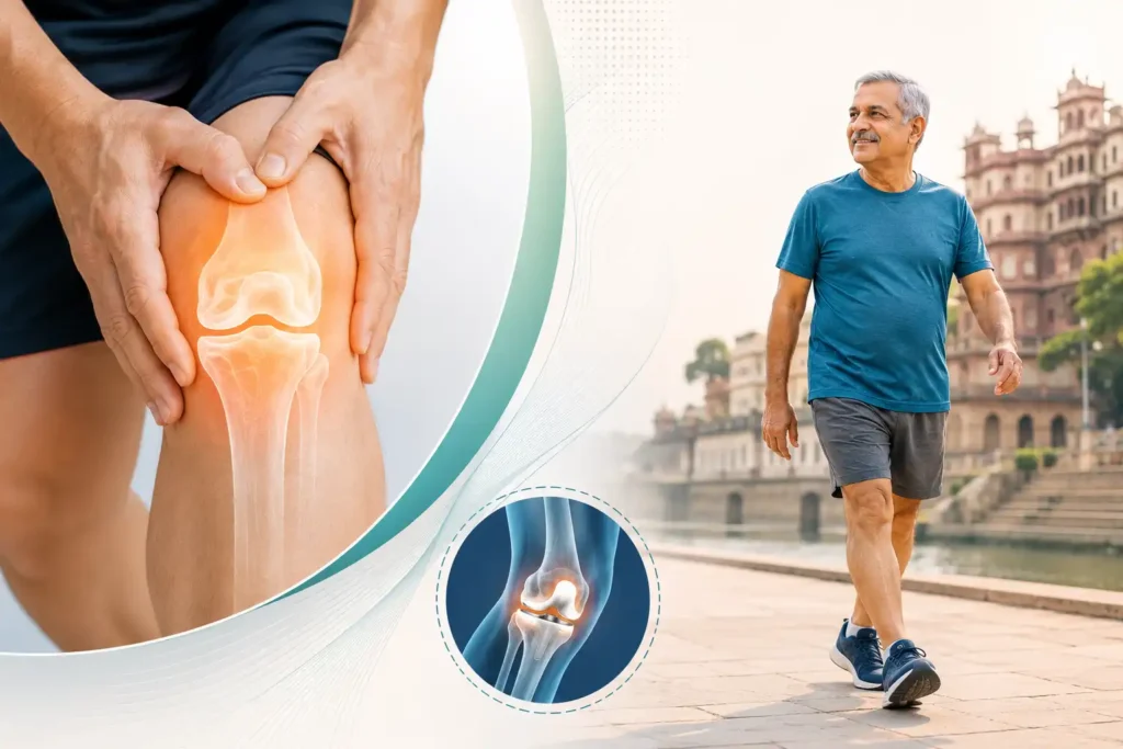
Every day at the clinic,
Dr. Prince Uchadiya meets at least one patient who says the same thing:
“Doctor, I have been having knee pain for years but everyone told me to just live with it until it gets bad enough for surgery.” Some of these patients are 55 and have Grade 2 osteoarthritis that could have been well managed with physiotherapy and lifestyle changes, preventing progression for years. Others are 70 with Grade 4 bone-on-bone disease who have waited so long that surgery is now the only path to pain relief. Both patients needed the same thing years earlier: an accurate understanding of which stage of the disease they were in and what the right treatment is for that specific stage.
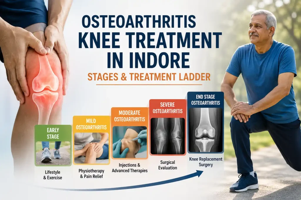
Knee osteoarthritis is not a single condition that you either have or you do not. It is a progressive disease with four radiographically defined stages, each with a different clinical picture, a different burden of pain and disability, and a different treatment approach. The concept of a treatment ladder, where increasingly intensive interventions are matched to increasing disease severity, is the most rational and evidence-based framework for managing knee osteoarthritis treatment in Indore. Understanding this ladder, and knowing where you sit on it, is the starting point for every patient who wants to take control of their knee health.
What Is Knee Osteoarthritis and How Does It Develop Over Time?
Knee osteoarthritis is the progressive degeneration of the articular cartilage that covers the bone surfaces within the knee joint, combined with secondary changes in the underlying bone, joint lining, ligaments, and periarticular muscles. The articular cartilage of a healthy knee is a smooth, glistening, shock-absorbing surface approximately five to seven millimetres thick that allows the femur and tibia to glide against each other with minimal friction during every movement. In osteoarthritis, this cartilage gradually breaks down. It loses its water content, its structural integrity, and its smooth surface. As the cartilage thins and becomes irregular, friction increases. The underlying bone reacts by thickening and developing bony projections called osteophytes at the joint margins. The joint space narrows progressively on imaging. The joint lining (synovium) produces inflammatory fluid in response to cartilage debris, producing the swelling and morning stiffness that are characteristic features of the condition.
Indian epidemiological data presents a striking picture: a multi-city study found that 28.7 percent of the adult population studied had radiographic knee osteoarthritis when assessed using the Kellgren-Lawrence grading scale. The condition is more prevalent in women than men (31.6 percent versus 25 percent in the study), and its prevalence increases sharply with age. Knee osteoarthritis is now recognised as one of the leading causes of disability in Indian adults over 60, affecting not just mobility but quality of life, independence, sleep, and mental health.
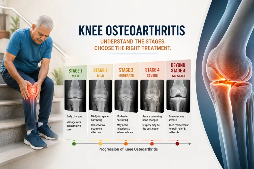
The Kellgren-Lawrence Staging System: How Knee Osteoarthritis Is Graded
The Kellgren-Lawrence (KL) grading scale is the universally accepted radiographic classification system for knee osteoarthritis severity. It assigns a grade from 0 to 4 based on specific findings seen on weight-bearing X-rays of the knee. Understanding these grades helps patients interpret what their X-ray report means in clinical terms and what treatment options are appropriate for each grade.
- Grade 0: No radiographic features of osteoarthritis. Normal knee. Joint space is well preserved, no osteophytes, no sclerosis.
- Grade 1 (Doubtful OA): Possible osteophytic lipping at the joint margins and doubtful joint space narrowing. The patient may have knee pain and symptoms, but the X-ray changes are minimal and borderline. This is the stage where intervention is most impactful for slowing progression.
- Grade 2 (Mild OA): Definite osteophytes present and possible joint space narrowing on weight-bearing X-ray. Cartilage loss is beginning. Morning stiffness, pain with activity, and occasional swelling are typical. Conservative treatment is highly effective at this stage.
- Grade 3 (Moderate OA): Multiple osteophytes, definite joint space narrowing, subchondral sclerosis (hardening of the bone beneath the cartilage), and possible bony deformity. Pain is more constant, activity is significantly limited, and stair climbing and prolonged walking become difficult. This is the stage where injections and more intensive physiotherapy come into play.
- Grade 4 (Severe OA): Large osteophytes, marked joint space narrowing (often to the point of bone-on-bone contact), severe sclerosis, and definite bony deformity. This is end-stage arthritis. Pain is constant, rest pain is present, and walking capacity is severely curtailed. Knee replacement surgery is the definitive treatment for Grade 4 disease when conservative management can no longer provide adequate quality of life.
Stage-by-Stage: The Knee Osteoarthritis Treatment Ladder in Indore
The treatment ladder concept is the most important framework for understanding knee osteoarthritis treatment in Indore. The principle is straightforward: treatment intensity and invasiveness increases in proportion to disease severity. Starting at the bottom rung with lifestyle modifications and physiotherapy and progressing upward only when each level fails to provide adequate relief is both scientifically supported and clinically rational. Applying knee replacement surgery to a patient with Grade 2 osteoarthritis who has never tried physiotherapy is as inappropriate as prescribing only painkillers to a patient with Grade 4 bone-on-bone disease who cannot walk to the bathroom.
Rung 1: Exercise, Weight Management, and Lifestyle Modification (Grade 1-2)
This is the most important rung of the treatment ladder and the one that is most consistently underutilised. For patients with Grade 1 and Grade 2 knee osteoarthritis, evidence-based clinical guidelines from international orthopaedic bodies unanimously identify exercise and weight management as the most effective interventions available. This is not a holding measure while waiting for the disease to worsen. It is the primary treatment that can meaningfully slow OA progression and maintain function for years.
The best-evidenced exercises for knee osteoarthritis are low-impact aerobic activities (swimming, cycling, walking on level ground), quadriceps strengthening exercises (straight leg raises, terminal knee extensions, mini squats at a pain-free range), and hip abductor strengthening. Strong quadriceps reduce the load transmitted to the articular cartilage with every step. Strong hip abductors reduce the varus (bow-leg) moment at the knee that concentrates load on the medial compartment, the most commonly affected area in Indian patients.
Weight is the most powerful modifiable risk factor for knee osteoarthritis progression. Each kilogram of body weight reduction produces an estimated four-kilogram reduction in knee joint load per step. In a patient who takes 7,000 steps per day, losing five kilograms reduces cumulative daily knee load by 140,000 kilograms. This is not a trivial clinical effect. Patients seeking knee osteoarthritis treatment in Indore who are overweight should be counselled that weight management is as important a treatment as any medication or injection.
Rung 2: Medications and Physiotherapy (Grade 2-3)
When pain levels are limiting the patient’s ability to participate in the exercise program, medications support the process by reducing pain sufficiently to allow activity. Anti-inflammatory medications such as ibuprofen or diclofenac are used for short-term pain reduction during flare-ups rather than as long-term daily therapy, due to gastrointestinal and cardiovascular risks with chronic use. Topical anti-inflammatories applied directly to the knee skin are effective for local pain relief with minimal systemic side effects.
Structured physiotherapy for Grade 2-3 osteoarthritis goes beyond simple home exercises. A physiotherapist assesses muscle imbalances, gait mechanics, and specific movement patterns that are overloading the arthritic compartment, and designs a program that simultaneously builds strength, reduces provocative loading, and educates the patient about joint protection techniques for daily activities. Patients in Indore can access structured rehabilitation through the post-injury rehabilitation program.
Knee braces and supports play a useful role for some patients. An unloading brace, which shifts the load away from the most affected knee compartment, can reduce pain during walking and allow higher activity levels. Simple sleeves provide proprioceptive feedback and warmth that reduces pain in many patients, though they do not alter the underlying disease.
Rung 3: Injections and Minimally Invasive Interventions (Grade 3)
When exercise, physiotherapy, and medications have been appropriately pursued but pain is limiting quality of life at Grade 3 severity, intra-articular injections are the next step. These deliver a therapeutic agent directly into the joint space, targeting the source of pain more precisely than systemic medications.
Corticosteroid injections reduce the inflammatory component within the arthritic joint and provide meaningful short-term pain relief typically lasting four to twelve weeks. They are particularly effective for Grade 3 osteoarthritis with significant joint effusion (swelling), and can provide a window of reduced pain during which physiotherapy can be more aggressively pursued. Multiple injections can be given but are spaced at intervals of at least three to four months to minimise the risk of cartilage effects from repeated steroid exposure.
Hyaluronic acid injections (viscosupplementation) deliver a gel-like substance that temporarily improves joint lubrication and reduces mechanical friction within the osteoarthritic joint. Evidence for hyaluronic acid is more variable than for corticosteroids in terms of immediate pain relief, but some studies show more sustained benefit at six months, making it a useful option for patients who need longer-term relief between interventions.
Platelet-Rich Plasma (PRP) injections deliver concentrated growth factors from the patient’s own blood into the arthritic joint, targeting the biological repair and anti-inflammatory pathways within the cartilage and synovium. Published randomised controlled trial evidence suggests that PRP injections produce superior outcomes to hyaluronic acid at six and twelve months in Grade 2-3 knee osteoarthritis, with particularly good results in younger patients (under 60) and those with earlier-stage disease. Information about PRP and regenerative injection options in Indore is available on the PRP versus stem cell treatment page.
Rung 4: Knee Replacement Surgery (Grade 4 and Refractory Grade 3)
Total knee replacement surgery is the most effective treatment for Grade 4 knee osteoarthritis and is also appropriate for selected Grade 3 patients who have failed all conservative and injection-based treatments and whose quality of life is significantly impaired despite appropriate management. The procedure replaces the damaged articular surfaces of the femur and tibia with precisely engineered metal and polyethylene components that restore pain-free joint mechanics.
Published data consistently shows that total knee replacement produces significant reduction in pain, meaningful improvement in walking capacity and quality of life, and high patient satisfaction rates exceeding 85 to 90 percent in appropriately selected patients. The surgery is not recommended for patients who have not adequately trialled conservative management, regardless of X-ray grade, because some patients with Grade 3 or even Grade 4 X-ray changes have manageable symptoms that do not justify the risks and recovery demands of surgery.
For eligible patients in Indore and Madhya Pradesh, knee replacement surgery can be accessed under the Ayushman Bharat PMJAY scheme and other government orthopaedic programmes. Detailed information is available on the Ayushman Bharat knee replacement Indore page and the general knee replacement surgery page.
Can Knee Osteoarthritis Happen Before 50? The Younger Patient Problem
Knee osteoarthritis is predominantly a condition of older adults, but it increasingly presents in patients in their 40s and even late 30s in Indore. Post-traumatic osteoarthritis, developing after ACL tears, meniscus injuries, or significant cartilage damage that was not adequately managed, accounts for a substantial proportion of younger OA patients. Elevated BMI, which accelerates cartilage wear under mechanical overload, is a significant driver of early-onset OA. Occupational factors including prolonged squatting, kneeling, and heavy lifting also contribute to premature joint degeneration in working-age adults.
Young patients with knee osteoarthritis present a specific treatment challenge because the goal is not just managing current symptoms but preserving the joint for as long as possible. For young patients with Grade 1-2 OA, the treatment focus is aggressively on weight management, quadriceps strengthening, activity modification, and cartilage-supportive measures including nutritional supplementation. Information about early cartilage changes and their management in Indore is available on the early cartilage damage page.
Lifestyle Habits That Slow Knee Osteoarthritis Progression
The trajectory of knee osteoarthritis is not fixed. While the underlying disease cannot currently be reversed, its rate of progression is significantly influenced by daily habits that are within the patient’s control. Consistent low-impact exercise that maintains quadriceps and hip strength without high-impact loading is the most effective single habit for slowing progression and maintaining function. Maintaining a healthy body weight removes the most significant modifiable mechanical load from the joint. Avoiding prolonged deep squatting and kneeling, which are particularly high-stress positions for the medial compartment, reduces cumulative daily cartilage loading. Using appropriate footwear with shock-absorbing insoles reduces impact loading with every step.
Dietary factors including adequate calcium and vitamin D intake support bone health and reduce the secondary bone changes associated with progressing OA. Omega-3 fatty acids have some evidence for modest anti-inflammatory benefit in osteoarthritis. Indian patients in Indore who are vegetarian should be aware of their vitamin D status, as deficiency is extremely common and can exacerbate musculoskeletal pain independent of the structural OA changes. Supplement use should be discussed with the treating specialist to ensure it is appropriate for the individual’s stage of disease and overall health.
Frequently Asked Questions About Knee Osteoarthritis Treatment Indore
1. What is knee osteoarthritis and how does it develop over time?
Knee osteoarthritis is the progressive degeneration of the articular cartilage covering the bone surfaces within the knee joint, combined with secondary changes in the underlying bone (sclerosis and osteophyte formation), joint lining, and periarticular soft tissues. It develops when the mechanical load on the joint, over years and decades, exceeds the cartilage’s capacity for self-repair. Risk factors including age, elevated body weight, previous knee injuries, occupational loading, and genetic predisposition all accelerate the process. The cartilage progressively thins and loses its smooth surface, joint friction increases, inflammation in the joint lining is triggered by cartilage debris, and bony spurs develop at the joint margins. The disease progresses gradually through four radiographically defined stages over years to decades.
2. What are the different stages of knee osteoarthritis?
Knee osteoarthritis is staged using the Kellgren-Lawrence grading scale on a spectrum from Grade 0 (normal) to Grade 4 (severe). Grade 1 (doubtful OA) shows possible osteophytes and borderline joint space changes. Grade 2 (mild OA) shows definite osteophytes and possible joint space narrowing. Grade 3 (moderate OA) shows multiple osteophytes, definite joint space narrowing, and subchondral sclerosis. Grade 4 (severe OA) shows large osteophytes, marked joint space narrowing approaching bone-on-bone contact, severe sclerosis, and bony deformity. Each grade corresponds to a different level of symptoms and a different appropriate treatment approach in the knee osteoarthritis treatment ladder.
3. How do doctors determine the severity or stage of knee osteoarthritis?
Stage is determined through a combination of clinical assessment and radiographic imaging. Standing weight-bearing X-rays of both knees, taken in the anteroposterior, lateral, and skyline views, are the standard investigation for OA grading. The KL grade is assigned based on the presence and extent of osteophytes, the degree of joint space narrowing, subchondral sclerosis, and bony deformity. Clinical findings including range of motion, joint line tenderness, the presence of swelling, and functional limitations are assessed alongside the X-ray to determine the overall clinical burden. MRI is used in specific situations where soft tissue detail is needed, such as to assess associated meniscal or cartilage damage in earlier-stage disease where surgical intervention for cartilage pathology might be considered.
4. Can knee osteoarthritis happen before age 50?
Yes, and it is increasingly common in patients in their late 30s and 40s in Indore. Post-traumatic arthritis developing after ACL tears, meniscus injuries, or significant cartilage damage is a major cause of early-onset OA. Elevated body weight accelerates cartilage degeneration under increased mechanical load. Occupational activities involving prolonged squatting, kneeling, and heavy lifting in younger workers cause premature cartilage wear. The treatment approach in younger patients prioritises joint preservation over symptom management, with aggressive conservative treatment aimed at maximising the functional lifespan of the native joint before replacement becomes necessary.
5. Why does knee osteoarthritis sometimes cause thigh pain or leg pain?
Knee osteoarthritis primarily produces pain at the knee joint itself, but referred pain extending into the thigh, calf, or even the hip is a recognised clinical feature. The reason is partly that the inflamed joint capsule and synovium produce pain signals that the nervous system maps imprecisely, causing the perceived pain to radiate beyond the anatomical joint location. Secondary changes in gait mechanics, where the patient alters their walking pattern to protect the painful knee, can also produce myofascial pain and muscle fatigue in the thigh and calf. Additionally, osteoarthritis often coexists with trochanteric bursitis, knee effusion-related quadriceps inhibition, and in older patients with concurrent lumbar spine or hip pathology, all of which contribute to a broader pain distribution than the knee alone.
6. What are the early warning signs of knee osteoarthritis?
Early warning signs of knee osteoarthritis that patients in Indore should recognise include morning stiffness lasting fifteen to thirty minutes that gradually resolves with movement, a cracking or crunching sensation (crepitus) felt or heard during knee movement, pain that is worse after prolonged activity and improves with rest, difficulty going down stairs compared to climbing them, mild swelling around the knee that appears after activity and resolves with rest, and localised tenderness along the inner joint line of the knee. These early signs, particularly when occurring in a patient over 45 with risk factors such as elevated body weight or previous knee injury, warrant an orthopaedic evaluation and standing X-rays to determine the KL grade and begin appropriate early intervention.
7. Can exercises help reduce osteoarthritis knee pain?
Yes. Published clinical guidelines from major orthopaedic bodies internationally designate exercise as a core, mandatory treatment for all stages of knee osteoarthritis, regardless of severity. The mechanism is multifactorial: quadriceps strengthening reduces joint loading by absorbing a greater proportion of compressive force during weight-bearing, hip strengthening reduces the varus moment that concentrates load on the medial compartment, aerobic exercise reduces systemic inflammation and supports weight management, and the neuromuscular improvements from exercise improve joint proprioception and reduce the risk of provocative mechanical events. Exercise should be appropriately guided for each patient’s OA stage and functional capacity.
8. Which exercises are considered helpful for knee osteoarthritis patients?
The most evidence-supported exercises for knee osteoarthritis are low-impact aerobic activities including cycling on a stationary or outdoor bicycle, swimming and water aerobics, and walking on level ground with appropriate footwear. Specific muscle-strengthening exercises include straight leg raises (performed with the knee fully extended to avoid joint compression), mini squats to a pain-free range, terminal knee extensions using a resistance band, and seated knee extensions and flexions. Hip abductor strengthening with resistance bands reduces medial compartment loading during walking. High-impact activities including running, jumping, and stair-climbing with heavy loads should be avoided or minimised during symptomatic flare-ups. The exercise program should be designed by a physiotherapist based on the individual patient’s strength, flexibility, and pain levels.
9. Can osteoarthritis knee pain improve without surgery?
Yes, for Grade 1, 2, and in many cases Grade 3 knee osteoarthritis, meaningful and sustained pain improvement is achievable with conservative treatment. Exercise, weight management, physiotherapy, activity modification, appropriate analgesic medications, and intra-articular injections collectively provide substantial pain relief and functional improvement that can be maintained for years with appropriate ongoing management. Surgery is not appropriate as a first-line treatment for any stage of knee OA, and Grade 4 patients with low symptom burden who function adequately can still be managed conservatively for significant periods. Surgery becomes the right choice when conservative management can no longer maintain adequate quality of life, not simply because X-ray changes are present.
10. What is the treatment ladder for osteoarthritis knee management?
The knee osteoarthritis treatment ladder follows the disease severity grading. Grade 1-2 OA is managed with exercise, weight reduction, activity modification, patient education, and in symptomatic cases short courses of anti-inflammatory medications. Grade 2-3 OA adds structured physiotherapy, topical analgesics, knee bracing when appropriate, and considers viscosupplementation or PRP injection for patients with inadequate symptom control. Grade 3 OA with significant pain and functional limitation is the primary indication for corticosteroid or PRP injection alongside ongoing rehabilitation. Grade 4 OA that has not responded to conservative management and that is significantly impairing daily life is the indication for knee replacement surgery. Each rung is tried and genuinely pursued before the next is considered.
11. When do orthopedic doctors recommend injections for knee osteoarthritis?
Injections are recommended for knee osteoarthritis treatment in Indore when Grade 2-3 disease is producing pain and functional limitation that is not adequately controlled by exercise, physiotherapy, and oral medications. They are also appropriate when a patient needs a period of reduced pain to participate more effectively in a physiotherapy program. Corticosteroid injections work best for acutely inflamed, swollen knees and when rapid relief is needed. PRP injections are preferred for younger patients, those with Grade 2-3 OA who want a longer-lasting biological intervention, and those who have had limited benefit from or prefer to avoid repeated steroids. Viscosupplementation (hyaluronic acid) is appropriate for moderate OA with a greater mechanical than inflammatory component.
12. When does knee osteoarthritis become severe enough for knee replacement surgery?
Knee replacement surgery is appropriate when Grade 4 osteoarthritis, or occasionally severe Grade 3 with significant functional limitation, has failed to respond adequately to appropriately trialled conservative management and is causing persistent pain that significantly impairs quality of life, sleep, independence, or daily activities. The decision is driven by the patient’s symptoms and functional status rather than the X-ray grade alone. A patient with Grade 4 on X-ray but manageable symptoms who is functioning adequately is not necessarily a surgical candidate. A patient with Grade 3 on X-ray but whose knee pain prevents walking beyond 100 metres, severely disrupts sleep, and has failed three to six months of structured conservative treatment, is a surgical candidate.
13. Can lifestyle changes slow down knee osteoarthritis progression?
Yes, and this is one of the most important messages for patients with early to moderate knee OA. Weight reduction meaningfully reduces the mechanical load on the articular cartilage with every step. Consistent low-impact exercise maintains the muscular support that protects the joint and has evidence for reducing the rate of cartilage loss in early OA. Avoiding high-impact activities and sustained deep squatting reduces daily cartilage stress. Optimising vitamin D levels, which are frequently deficient in Indian adults, supports bone and cartilage health. These lifestyle measures cannot reverse established cartilage loss, but they reliably slow the progression of Grade 1-2 OA and delay the need for more intensive interventions at higher grades.
14. When should someone in Indore see an orthopedic doctor for osteoarthritis knee pain?
You should seek knee osteoarthritis treatment in Indore from a specialist when knee pain has been present for more than six weeks, when morning stiffness consistently takes more than thirty minutes to resolve, when knee swelling recurs repeatedly after activity, when you notice progressive difficulty with stairs, prolonged walking, or getting up from low chairs, when pain is beginning to affect sleep quality, or when you are self-managing with daily painkillers without an established diagnosis. Earlier evaluation allows the OA grade to be determined, appropriate treatment to be started for that grade, and a management plan to be established that gives the joint the best chance of preserved function for as long as possible. Waiting until the knee is bone-on-bone before seeking advice narrows the treatment options significantly.
15. What treatment options are available for osteoarthritis knee treatment in Indore?
Knee osteoarthritis treatment in Indore at Dr. Prince Uchadiya’s clinic covers the full treatment ladder. For early-stage disease, structured physiotherapy with a specific knee OA exercise prescription, weight management counselling, and activity modification guidance are provided. For Grade 2-3 disease, targeted physiotherapy, oral and topical analgesics, and intra-articular injections including corticosteroid, PRP, and viscosupplementation are available. For Grade 4 disease that has failed conservative management, knee replacement surgery is performed with Ayushman Bharat PMJAY coverage available for eligible patients. Every patient receives a treatment plan based on their KL grade, symptom severity, functional goals, and individual medical profile, with a clearly explained pathway of what comes next if the current treatment level does not produce adequate improvement.
If you have been managing knee pain with painkillers and hoping it will resolve on its own, the most important step you can take today is to find out which stage your osteoarthritis is at and start the right treatment for that stage. Knee osteoarthritis treatment in Indore at Dr. Prince Uchadiya’s clinic begins with a clear diagnosis and a treatment plan designed around where you are in the disease, not where you fear you might be heading. Book your consultation today.