Key Takeaways: From Dr. Prince Uchadiya’s Clinical Experience in Indore
- Clubfoot affects 1 in every 1,000 live births globally, making it one of the most common congenital musculoskeletal deformities. 80% of all clubfoot cases are born in low and middle-income countries including India. PMC India CTEV Study
- The biggest mistake I see families make is waiting too long because the child is “not complaining of pain.” A newborn with untreated clubfoot cannot tell you it hurts. The foot is being shaped every single week by its own soft tissue tension. The earlier treatment begins, the fewer casts needed, the lower the chance of surgery, and the better the long-term foot shape and function.
- The Ponseti method achieves full correction in over 94% of cases in India when started early and followed with consistent brace wear. Indian Tertiary Care Long-Term Outcomes Study, 2026
- Bracing is not optional after casting. It is the part most families stop early because the child seems fine after correction. Relapse almost always happens because the brace was abandoned. I tell every family: the casting corrects the foot, but the brace keeps it corrected for the years the foot is still growing.
- 66% of clubfoot cases in India are bilateral, meaning both feet are affected. Unilateral clubfoot affects the right foot more commonly than the left. PMC 2026
- Surgery is needed in a minority of cases, typically when treatment was started late, when the deformity is severe and rigid, or when relapse occurs after inadequate bracing compliance.
- Older children and adults with untreated clubfoot can still achieve significant functional improvement with treatment, though the approach shifts from casting to surgical correction of the underlying bony and soft tissue deformity.
- Clubfoot treatment in Indore at Dr. Prince Uchadiya Orthopaedic And Joint Care Clinic follows the Ponseti protocol with Pirani scoring, weekly cast changes, percutaneous Achilles tenotomy where required, and structured brace follow-up.
What Clubfoot Is and Why Early Clubfoot Treatment in Indore Matters
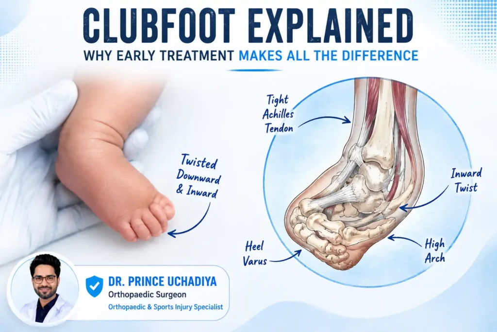
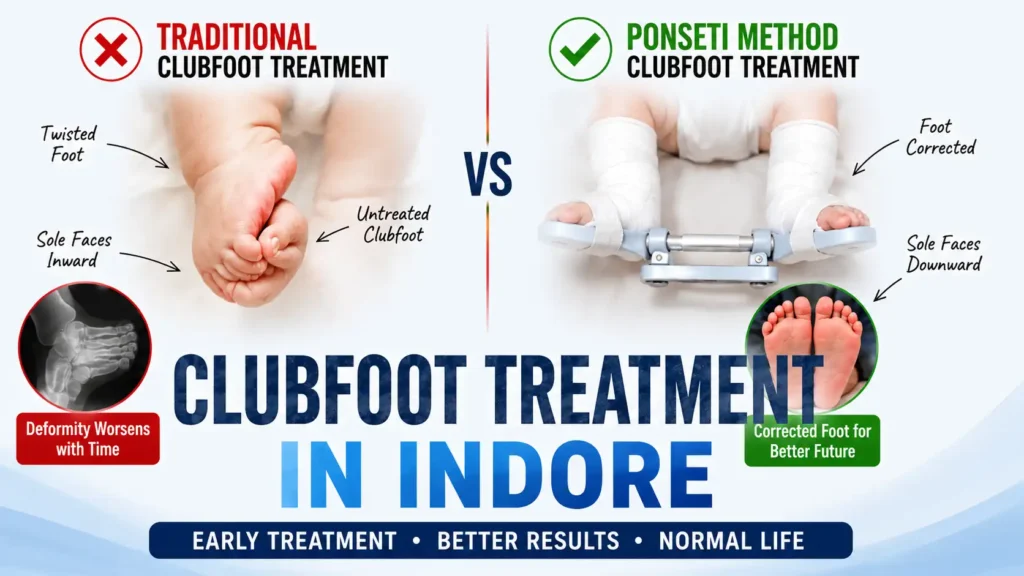
Clubfoot, medically known as Congenital Talipes Equinovarus or CTEV, is a structural deformity of the foot present at birth. The foot is twisted inward and downward, with the sole facing the other foot rather than the floor. It is not a skin or surface problem. The bones, tendons, ligaments, and muscles of the foot and ankle are all involved in a complex three-dimensional deformity that must be corrected systematically.
“When parents bring a newborn with clubfoot to the clinic, the first thing I explain is that this is a deformity, not a disease, and it is one of the most correctable conditions in orthopaedics when managed correctly from the start. The foot looks alarming, but the outcome with proper treatment is a completely functional, normal-looking foot. What makes the difference is timing and compliance.,” said Dr. Prince Uchadiya.
Left untreated, clubfoot does not resolve on its own. The child walks on the outer border or top of the foot, develops thick calluses, cannot wear normal footwear, and faces lifelong mobility limitations. In a study context relevant to Indore and central India, treatment delay is one of the most consistent factors linked to worse outcomes and higher surgical rates.
The Four Components of Clubfoot Deformity: CAVE
Every clubfoot has four deformity components that must be corrected in a specific sequence. Understanding these helps parents understand why each cast in the series is doing something different:
- C: Cavus (high arch): The arch of the foot is raised abnormally. This is corrected first by supinating the forefoot to align it with the hindfoot.
- A: Adductus (forefoot turned inward): The front of the foot points toward the other foot. Corrected during the early casting stages.
- V: Varus (heel turned inward): The heel tilts inward. Corrected progressively as the forefoot aligns.
- E: Equinus (foot pointed downward, heel raised): The most challenging component, requiring the Achilles tendon to be lengthened, usually through a minor percutaneous tenotomy.
The Pirani scoring system assigns a score of 0 to 6 based on these components, with higher scores indicating more severe deformity. This score is recorded at every visit to track correction progress objectively.
Who Gets Clubfoot: Risk Factors and Pattern in Indore
- Sex: Boys are affected approximately 3 times more often than girls
- Laterality: 66% of cases involve both feet; 34% are one-sided
- Family history: A parent with clubfoot increases the child’s risk significantly
- Associated conditions: Spina bifida, arthrogryposis, and certain neuromuscular conditions can cause clubfoot alongside the primary diagnosis
- Idiopathic: The majority of cases have no identifiable cause and occur in otherwise healthy infants
“In Indore, I see a consistent pattern of families arriving late, sometimes at 3 or 4 months when they should have been here at 2 to 3 weeks of age. The reason is usually that the treating doctor or the family did not realise how time-sensitive this is, or they waited to see if the foot would improve on its own. It will not improve on its own. Every week of delay in the newborn period makes the soft tissues stiffer and the correction harder.,” said Dr. Prince Uchadiya.
Diagnosis: How Clubfoot Is Identified and Scored
Clubfoot is frequently identified on antenatal ultrasound from around 20 weeks of pregnancy. This allows families to be counselled before birth and treatment to begin within the first week of life. When identified at birth, clinical examination confirms the diagnosis and the Pirani score is recorded.
The examination assesses:
- Hindfoot score (0 to 3): Posterior crease severity, heel shape emptiness, and rigidity of the equinus
- Midfoot score (0 to 3): Medial crease severity, talar head coverage, and lateral border curvature
- Total Pirani score: 0 to 6, with 6 being the most severe
X-rays are not routinely needed in newborns because the bones are mostly cartilage and not yet fully visible. In older children and adults presenting late, X-ray assessment of the talocalcaneal angle and foot alignment guides surgical planning.
Clubfoot Treatment in Indore: The Ponseti Method Step by Step
The Ponseti method is the globally recognised gold standard for clubfoot treatment in Indore and worldwide. It avoids extensive surgery in the vast majority of cases and produces a functional, pain-free, plantigrade foot that a person can walk on normally for life.
- Week 1 to 2: Assessment and first cast
The foot is gently manipulated and the first below-knee cast is applied, holding the correction achieved. No force is used. The foot is stretched to the point of gentle resistance only. The cast is moulded carefully around the heel and ankle. - Weeks 2 to 6: Weekly cast changes
The cast is changed weekly. At each change, the deformity components are corrected progressively in the CAVE sequence. The average number of casts needed in Indian studies is 5 to 7. Each cast holds the correction achieved that week and stretches slightly further. The Pirani score is re-recorded at each visit to track objective progress. - Achilles tenotomy for equinus correction
When all components except the equinus are corrected, the Achilles tendon is tight and is preventing the heel from coming down. A percutaneous tenotomy is performed under local anaesthesia in the clinic. A small needle-sized cut releases the tendon through the skin without any open incision. A final cast is applied for 3 weeks while the tendon heals. Indian data shows tenotomy is required in approximately 77% of CTEV feet. - Foot Abduction Orthosis (Denis-Browne brace)
After casting is complete, the corrected foot is held in a Denis-Browne brace. Both feet are attached to a bar that keeps the feet turned outward. For the affected foot, the angle is 60 to 70 degrees of external rotation. For the unaffected foot in unilateral cases, 30 to 40 degrees. The brace is worn 23 hours per day for the first 3 months, then nights and naps until age 4 to 5 years.
“The tenotomy sounds frightening to parents but it takes 10 seconds, the child cries briefly, and the relief in the foot position is immediate. What I spend most of my counselling time on is the brace, not the tenotomy. Parents accept the casting phase because the foot is visibly changing. But once the foot looks normal, they relax and stop the brace. That is when relapse happens.,” said Dr. Prince Uchadiya.
Ponseti Method vs Surgery: A Direct Comparison
| Factor | Ponseti Method | Surgical Correction |
|---|---|---|
| Age suitability | Newborn to approximately 2 years | Older children, rigid deformity, relapse cases |
| Invasiveness | Minimal: only percutaneous tenotomy | Open surgery: tendon releases, bony corrections |
| Success rate | Over 94% with compliance | Varies; higher complication risk |
| Recovery time | Functional within weeks of bracing | Months of recovery and rehabilitation |
| Stiffness risk | Low | Higher due to scar tissue formation |
| Relapse risk | Low with brace compliance | Possible; further surgery sometimes needed |
| Cost | Significantly lower | Significantly higher |
| Long-term foot quality | Supple, functional foot | Good but may be stiffer long-term |
When Surgery Becomes Necessary for Clubfoot Treatment in Indore
Surgery is not the failure of Ponseti. It is a different tool for a different situation. The specific scenarios where surgical intervention becomes part of the clubfoot treatment plan are:
- Resistant clubfoot: A small proportion of feet, typically those with the highest Pirani scores, do not achieve full correction with serial casting
- Late presentation: Children presenting after age 2 to 3 have stiffer soft tissues and bones that do not respond fully to casting alone
- Relapse after inadequate bracing: Recurrence of deformity when the brace was stopped early
- Neuromuscular clubfoot: Associated with spina bifida or cerebral palsy, where the underlying neurological imbalance causes ongoing deforming forces
- Older children and adults with untreated clubfoot: Require bony realignment procedures
The surgical procedures used in these situations include:
- Posteromedial soft tissue release: Lengthening of tight tendons and release of contracted ligaments on the inner and back of the foot. Used for younger children with resistant deformity
- Tibialis anterior tendon transfer: When the front outer part of the foot repeatedly supinates during walking after apparent correction, transferring this tendon to the middle of the foot corrects the muscle imbalance
- Calcaneal osteotomy: A cut in the heel bone to reposition it in the correct alignment, used in older children with persistent heel varus
- Triple arthrodesis: Fusion of the subtalar, talonavicular, and calcaneocuboid joints in adults or older adolescents with severe, rigid, uncorrected deformity. This eliminates painful motion at severely deformed joints and allows weight-bearing on a stable plantigrade foot
For patients needing any of these procedures, the fracture and trauma management experience at the clinic informs the surgical approach, and the minimally invasive surgery principles are applied wherever feasible to reduce recovery time.
Bracing Compliance: Why This Is the Most Important Part of Treatment
The correction achieved by casting is real. But the muscles and tendons that caused the deformity are still present. During the years of foot growth, if not held in the corrected position by the brace, those forces pull the foot back toward deformity. This is not a treatment failure. It is a predictable biological event that the brace prevents.
- Brace worn 23 hours per day for first 3 months after casting
- Then worn during all sleep hours until age 4 to 5 years
- Brace removed only for bathing and physiotherapy exercises
- Both feet are in the brace even if only one foot was affected
- The bar connecting the shoes must be rigid, not flexible
- The brace must fit correctly at every follow-up visit and be replaced as the child grows
“I have seen children with perfect casting outcomes at 3 months whose feet relapsed completely by age 2 because the brace was removed after a few weeks. And I have seen late-presenting children with severe Pirani scores achieve excellent long-term outcomes because the family was disciplined about the brace for 4 years. The brace is not a burden. It is the actual treatment.,” said Dr. Prince Uchadiya.
After the bracing phase, structured physiotherapy supports muscle balance and foot mechanics. The post-injury rehabilitation programme at the clinic provides guidance for families transitioning from the brace phase into active foot strengthening.
Clubfoot in Older Children and Adults: Is It Too Late?
Families sometimes arrive with older children who were never treated, or whose treatment was incomplete. The same question comes from adults who grew up walking on the outer border of their foot and have accepted it as their normal. The answer is that it is never too late to improve function significantly, though the approach and realistic outcome goals shift with age.
- Ages 2 to 5: Modified Ponseti casting still works in many cases, with more casts needed. Tibialis anterior tendon transfer is commonly added for residual supination
- Ages 5 to 12: Bony corrections become more central. Calcaneal osteotomies, midfoot osteotomies, and tendon procedures are combined based on the specific deformity pattern
- Adolescents and adults: Triple arthrodesis or selective joint fusions stabilise the foot in a functional position and eliminate chronic pain. The goal shifts from achieving perfect anatomy to achieving a foot that can bear weight comfortably and fit into footwear
For families in Indore with older children or adults seeking evaluation of untreated or inadequately treated clubfoot, a structured second opinion at the clinic provides a clear assessment of what correction is realistic and what the treatment pathway looks like at the current age and deformity severity.
What to Expect at the First Clubfoot Consultation in Indore
The first visit to Dr. Prince Uchadiya Orthopaedic And Joint Care Clinic in Nipania, Indore for a clubfoot assessment covers the following in a single appointment:
- Full clinical examination of both feet and overall lower limb alignment
- Pirani scoring of the deformity
- Assessment of any associated conditions including hip dysplasia, spinal anomalies, or neurological findings
- Clear explanation to parents of what the Ponseti sequence involves, week by week
- First cast applied at the same visit if the infant is appropriate age and the parents are counselled
- Follow-up schedule planned: weekly for the casting phase, then monthly through the bracing phase
Families from across Indore, including Vijay Nagar, Scheme 54, Mahalaxmi Nagar, Palasia, and from districts including Dewas, Ujjain, and Dhar, attend the Nipania clinic for clubfoot treatment in Indore. The structured follow-up protocol is designed to work around family logistics while maintaining the weekly frequency that the casting phase requires.
Frequently Asked Questions: Clubfoot Treatment in Indore
1. Can clubfoot be detected before birth?
Yes. Antenatal ultrasound from around 18 to 20 weeks of pregnancy can identify clubfoot. This allows the family to be counselled in advance, understand the treatment process before birth, and begin treatment in the first week of life rather than presenting weeks later after uncertainty and delay. Prenatal detection does not change the treatment but it significantly improves the timing of starting it.
2. What is the ideal age to start Ponseti treatment?
The first week of life is ideal. The soft tissues of a newborn are at their most pliable due to circulating maternal hormones, and the foot responds to gentle manipulation most rapidly at this stage. Treatment can still begin successfully up to 2 years of age with a similar Ponseti protocol, though more casts are needed and the response is slower. After 2 years, modified approaches and sometimes surgical correction are required.
3. How many casts will my child need?
The average in Indian studies is 5 to 7 casts for full correction. A mild Pirani score foot may correct in 4. A severe, stiff foot may need 8 or more. Each cast is changed weekly. After the casting series, a percutaneous Achilles tenotomy is performed in approximately 77% of cases, followed by a final 3-week cast while the tendon heals.
4. Does the percutaneous tenotomy hurt the baby?
The tenotomy is performed under local anaesthesia using a very fine needle. The Achilles tendon in a newborn is very small. The procedure takes seconds. The child cries briefly from the local anaesthetic injection and settles quickly. There is no open wound. A cast is applied immediately and the tendon heals fully over the following 3 weeks.
5. What happens if we miss a week of casting?
Missing a cast change by a few days during the active correction phase allows the soft tissues to begin reverting toward the deformed position. The correction from that cast is partially lost. Consistency with weekly appointments is not a recommendation. It is a clinical requirement. If an appointment must be missed, contact the clinic immediately to reschedule within the same week rather than waiting for the next scheduled slot.
6. My child’s foot looks completely normal now after casting. Can we stop the brace?
No. The foot looking normal after casting is the intended result of casting. But the deforming forces in the tendons and muscles are still present. Without the brace to hold the foot in correction during the years of growth, relapse is highly likely. The brace is not treating the foot that looks wrong. It is preventing the foot that looks right from relapsing. This is the most important message in the entire treatment programme.
7. Can a child with clubfoot play sports and live a normal life?
Yes. Children who complete the full Ponseti protocol with consistent bracing grow up with functional, pain-free feet. They walk normally, run, play sports, and wear standard footwear. Long-term studies show that adults who were treated with the Ponseti method in infancy report minimal functional limitations compared to the general population. The goal of treatment is a completely functional life, not just correction of appearance.
8. Is clubfoot related to anything I did during pregnancy?
No. Clubfoot is not caused by anything a mother did or did not do during pregnancy. It is a complex developmental deformity with genetic and environmental factors that are not under the mother’s control. Families sometimes carry guilt about this unnecessarily. The condition occurs in otherwise healthy pregnancies with no complications, and its cause in most cases is genuinely unknown.
9. My child is 4 years old and has clubfoot that was never properly treated. What can be done?
At 4 years, modified casting may still play a role for some components of the deformity, but surgical correction of the bony and soft tissue elements is likely needed. The specific procedures depend on the deformity pattern seen on clinical examination and X-ray. Correction at this age is more complex than in infancy but still achieves significant functional improvement. A consultation at the clinic will define exactly what is possible and what the treatment timeline looks like for the specific severity of deformity present.
10. Is clubfoot treatment covered under Ayushman Bharat in Indore?
Certain clubfoot treatment procedures, including casting-related care and surgical correction procedures, fall within PM-JAY coverage for eligible patients. The specific package applicable depends on the procedure required. Patients with Ayushman cards should discuss eligibility and procedure coverage at the time of consultation. The clinic’s experience with the Ayushman Bharat surgical process includes guiding families through eligibility confirmation and pre-authorization for paediatric orthopaedic procedures.