Twenty years ago, repairing a torn knee ligament, stitching a detached shoulder tendon, or removing a loose fragment of bone meant a 10-to-15-centimeter open surgical incision, cutting through healthy muscle layers, days of severe post-operative pain, and weeks in a heavy plaster cast. Today, over 90% of internal sports, ligament, meniscus, labral, and early cartilage procedures are performed through tiny 4mm to 5mm keyhole incisions no wider than a buttonhole.
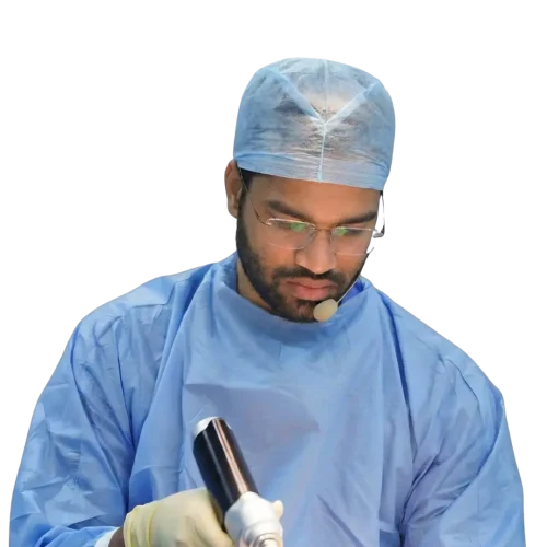
This minimally invasive technique is called arthroscopic surgery (joint keyhole surgery). At Dr. Prince Uchadiya Orthopaedic & Joint Care Clinic, Dr. Prince Uchadiya (MS Orthopaedics Gold Medalist from KEM Hospital Mumbai, MRCS from the Royal College of Surgeons of England, with 500+ arthroscopic surgeries performed) uses high-definition 4K fiber-optic arthroscopy to diagnose and repair complex knee, shoulder, ankle, and hip injuries with minimal tissue trauma and rapid functional recovery.
What Is Arthroscopic Surgery and How Does Keyhole Joint Repair Work?
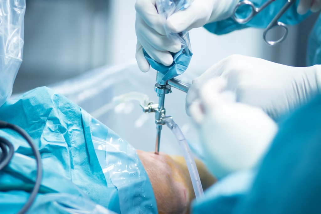
The word arthroscopy comes from two Greek words: arthro (joint) and skopein (to look inside). According to the American Academy of Orthopaedic Surgeons (AAOS OrthoInfo), an arthroscope is a slender, pencil-sized metal tube containing a system of precision lenses and fiber-optic lighting connected to a high-definition 4K surgical monitor.
Instead of slicing open the joint capsule and retracting muscles to see inside with the naked eye, the orthopaedic surgeon creates two or three tiny 5-millimeter portals in the skin:
- Sterile Saline Joint Expansion: Sterile physiological fluid is gently pumped through the joint cavity. This expands the tight joint space, washes away cloudy inflammatory fluid or blood, and gives the surgeon a crystal-clear, magnified 360-degree view of every ligament, cartilage surface, and tendon attachment.
- Magnified Dynamic Inspection via Viewing Portal: Because the 4K camera magnifies internal structures 10 to 15 times on the operating monitor and has a 30-degree or 70-degree angled lens, the surgeon can look around corners (such as the posterior horn of the meniscus or the undersurface of the rotator cuff) that are virtually impossible to see even during open surgery.
- Precision Repair via Working Portals: Through a second 5mm keyhole, specialized micro-instruments (motorized arthroscopic shavers, radiofrequency coblation wands, all-inside meniscal suture passers, and bio-absorbable suture anchors) are introduced to stitch torn tissue, reconstruct ligaments, or smooth damaged cartilage.
Keyhole Arthroscopic Surgery vs Traditional Open Joint Surgery
Clinical guidelines from Mayo Clinic and Johns Hopkins Medicine highlight why keyhole arthroscopy has replaced open arthrotomy for almost all soft-tissue joint reconstructions:
| Surgical Parameter | Keyhole Arthroscopic Surgery | Traditional Open Joint Surgery (Arthrotomy) |
|---|---|---|
| Incision Size | Two or three 4mm to 5mm keyholes (closed with 1 stitch each) | 8 cm to 15 cm full skin and joint capsule incision |
| Muscle & Capsule Trauma | Micro-instruments pass gently between muscle fibers without cutting them | Requires splitting or detaching muscles and opening the entire joint capsule |
| Intra-Operative Visualization | 10x-15x 4K magnification; reaches deep posterior corners of knee and shoulder | Limited line-of-sight view; deep posterior structures are difficult to access |
| Blood Loss & Infection Risk | Virtually bloodless (<10-20 mL); continuous sterile saline irrigation keeps infection risk <0.2% | Higher blood loss and longer wound exposure to air |
| Post-Operative Pain & Scar Tissue | Mild soreness managed with oral tablets; minimal internal scar tissue (arthrofibrosis) | Significant pain during Weeks 1-2; higher risk of post-op joint stiffness |
| Hospital Stay & Walking | Day-care or 1 overnight stay; walking begins the very next morning | 3 to 5 nights hospital stay; slower mobilization |
Conditions Treated with Keyhole Arthroscopy at Dr. Prince Uchadiya’s Clinic
On our dedicated arthroscopy surgery service page, we categorize keyhole procedures by the specific joint involved:
1. Knee Arthroscopy (Ligaments, Meniscus & Cartilage)
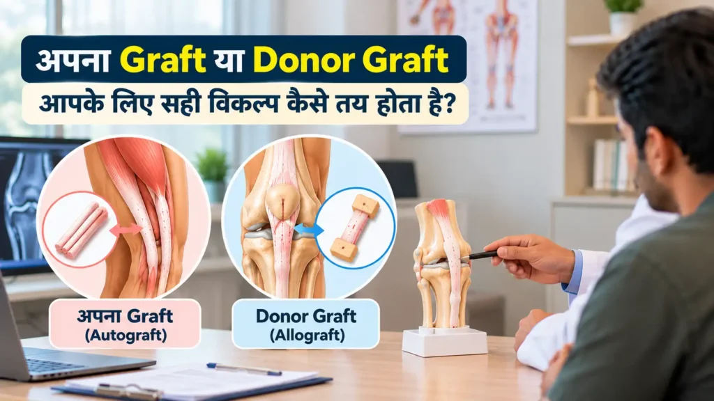
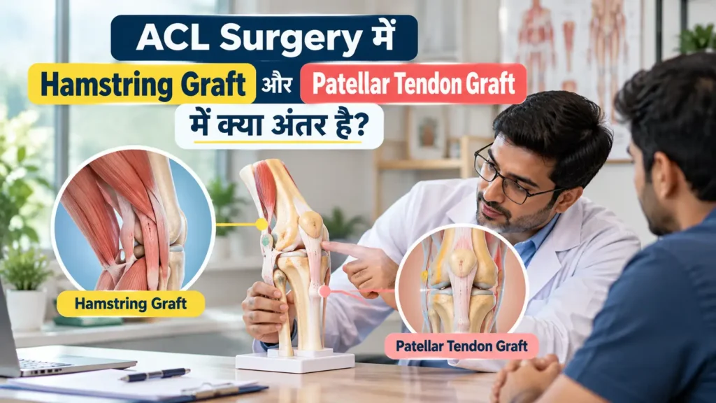
- Anatomical ACL & PCL Reconstruction: Replacing a torn cruciate ligament using Peroneus Longus, Hamstring, or BTB autografts fixed with cortical buttons and biocomposite screws. Read our complete guide on ACL reconstruction surgery in Indore.
- All-Inside Meniscus Repair & Root Fixation: Stitching bucket-handle, radial, or root tears of the shock-absorbing meniscus cushion, especially in combined ACL and meniscus injuries or Grade 3 meniscus tears.
- Focal Articular Cartilage Restoration: Treating early and focal cartilage defects via arthroscopic microfracture, chondroplasty, or Osteochondral Autograft Transfer (OATS / Mosaicplasty).
- Loose Body Removal & Synovectomy: Extracting floating bone/cartilage fragments that cause sudden knee locking, or clearing inflamed synovium in rheumatoid or pigmented villonodular synovitis.
2. Shoulder Arthroscopy (Rotator Cuff & Recurrent Dislocation)
- Double-Row Arthroscopic Rotator Cuff Repair: Re-anchoring torn supraspinatus and infraspinatus tendons back to the humeral footprint using high-strength biocomposite suture anchors. See our guide on shoulder rotator cuff tear treatment in Indore.
- Bankart Labral Repair & Remplissage: Re-attaching the torn glenoid labrum bumper and filling Hill-Sachs defects to stop recurrent shoulder dislocations in young athletes.
- Subacromial Decompression & Capsular Release: Shaving down a hooked acromion bone spur pinching the tendon, or releasing a thick, contracted joint capsule in refractory frozen shoulder (adhesive capsulitis).
3. Ankle, Hip & Elbow Arthroscopy
- Ankle Arthroscopy: Removing anterior/posterior bone impingement spurs in footballers and runners, and treating osteochondral defects of the talus.
- Hip Arthroscopy: Treating femoroacetabular impingement (FAI) cam/pincer bone bumps and acetabular labral tears in young adults.
| Arthroscopic Procedure | Typical OT Duration | Hospital Stay | Desk Work Return | Full Sports / Heavy Load Return |
|---|---|---|---|---|
| Partial Meniscectomy / Loose Body Removal | 25 to 40 minutes | Day-care / 1 night | 5 to 7 days | 4 to 6 weeks |
| All-Inside Meniscus Suture Repair | 40 to 60 minutes | 1 night | 7 to 14 days | 4 to 6 months |
| Anatomical ACL Reconstruction | 45 to 75 minutes | 1 to 2 nights | 10 to 14 days | 6 to 9 months (with milestone rehab) |
| Arthroscopic Bankart Repair (Shoulder Dislocation) | 45 to 60 minutes | 1 night | 10 to 14 days (with sling) | 4 to 6 months |
| Arthroscopic Rotator Cuff Repair | 60 to 90 minutes | 1 to 2 nights | 2 to 3 weeks | 5 to 6 months |
Preparing for Surgery and Mastering Post-Arthroscopy Rehabilitation
Because keyhole incisions heal rapidly on the outside, some patients mistakenly assume the deep biological repair inside the joint is also healed in two weeks. To protect your suture anchors or ligament graft, we coordinate every surgery with a structured post-arthroscopy physiotherapy protocol, combining supervised clinic and home physiotherapy, evidence-based cartilage and tendon nutrition, and long-term conditioning to keep your joints strong after surgery and prevent future sports injuries.
For complete package estimates and cashless insurance details in Indore, read our transparent guide on arthroscopy surgery cost in Indore.
Step-by-Step Patient Journey on the Day of Arthroscopic Surgery in Indore
For many patients and families, fear of the unknown is the biggest barrier to fixing a torn ligament or meniscus. Knowing exactly what happens from hospital admission to next-morning discharge removes that anxiety completely:
- Pre-Anaesthesia Checkup & MRI Mapping (1 to 3 Days Prior): Once your clinical exam and 1.5T/3T MRI confirm a surgical tear, a routine blood panel, ECG, and anaesthesiologist fitness check are completed. Our insurance desk submits your cashless pre-authorization to your TPA so paperwork is cleared before admission.
- Morning Admission & Regional Nerve Block: You arrive at the hospital fasting on the morning of surgery. Inside the modular Laminar-Airflow OT, the anaesthesiologist administers a comfortable spinal block (for knee/ankle) or ultrasound-guided regional nerve block (for shoulder) so your limb is completely numb and pain-free.
- 45-to-60-Minute 4K Keyhole Procedure: Through two 5mm portals, Dr. Prince Uchadiya performs the anatomical repair using imported medical-grade implants (Smith & Nephew, Arthrex, Stryker, or Zimmer Biomet). High-definition intra-operative photos and videos are recorded for your medical file.
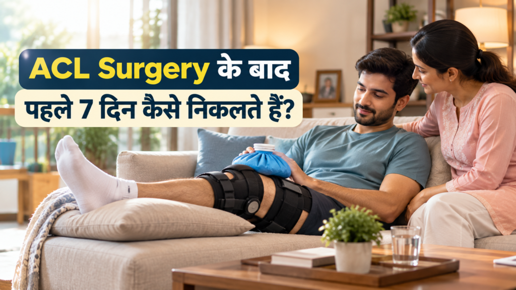
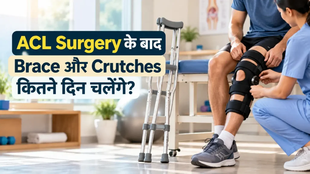
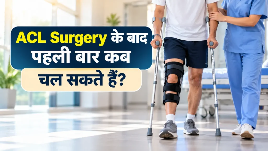
- Same-Evening Ankle Pumps & Next-Morning Assisted Walking: Within 3 to 4 hours of surgery, you are sitting up in your room sipping fluids and performing ankle pumps. The next morning, our physiotherapist guides you to stand and walk with a brace/walker, teaches your home exercise routine, and you return home with waterproof portal dressings and just two single-stitch keyholes.
Why Surgeon Fellowship Training & 4K Optical Precision Matter in Arthroscopy
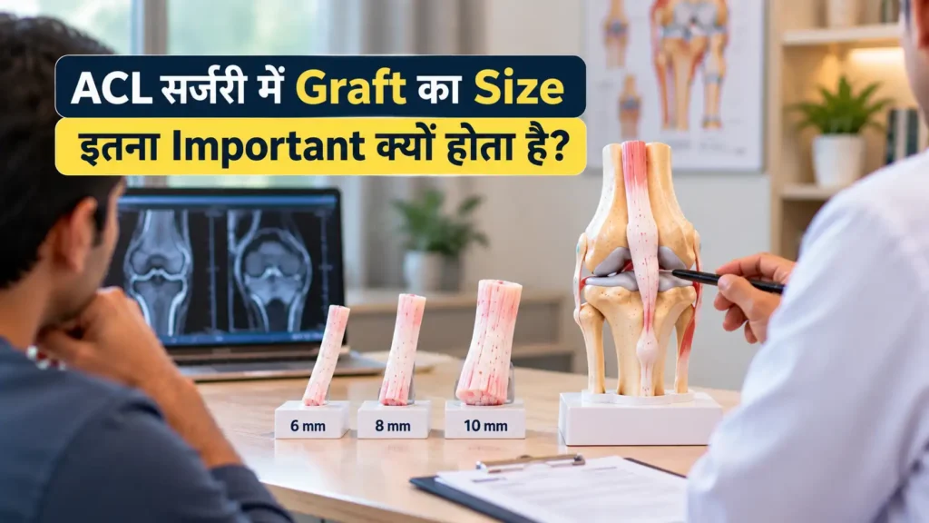
Because arthroscopic surgery is performed while looking at a 2D/4K monitor while manipulating micro-instruments in 3D space inside a fluid-filled cavity, hand-eye triangulation skill and anatomical tunnel accuracy make 100% of the difference between a joint that feels completely natural and one that feels tight or unstable. A tunnel placed just 3 millimeters too far forward during ACL reconstruction can restrict knee bending or cause graft impingement.
With Gold Medalist MS Orthopaedics training from KEM Hospital Mumbai, MRCS from the Royal College of Surgeons of England, and over 500+ arthroscopic reconstructions performed, Dr. Prince Uchadiya combines anatomical footprint preservation, all-inside meniscal saving techniques, and transparent implant documentation for every patient across Indore and Madhya Pradesh.
Frequently Asked Questions About Arthroscopic Surgery in Indore
1. Is arthroscopic keyhole surgery painful?
Knee and ankle arthroscopies are performed under comfortable spinal regional anaesthesia, while shoulder arthroscopies use general anaesthesia paired with an ultrasound-guided interscalene nerve block. You feel zero pain during the procedure, and because no major muscles are cut, post-operative discomfort in the first 48 hours is mild and easily controlled with oral medications and ice packs.
2. How big are the scars after knee or shoulder arthroscopy?
Each portal incision is only 4mm to 5mm long (about the width of a grain of rice) and requires just a single fine suture. Within 3 to 6 months, the tiny marks fade into almost invisible skin lines.
3. How soon can I walk and drive after knee arthroscopy?
After simple arthroscopic procedures (loose body removal, chondroplasty, or partial meniscectomy) and isolated ACL reconstruction, you stand and walk the very next morning. Most patients resume driving a car or two-wheeler within 2 to 4 weeks depending on the exact repair. See our guide on when you can safely drive after knee arthroscopy.
4. Can severe Grade 4 bone-on-bone knee osteoarthritis be cured with arthroscopy?
No. Clinical trials show that “washing out” a severely arthritic Grade 4 bone-on-bone knee does not regrow lost cartilage. Arthroscopy is highly effective for mechanical tears (ACL, meniscus, focal cartilage craters, loose bodies) in preserved joints, whereas advanced Grade 4 arthritis is treated with alignment osteotomy or muscle-sparing knee replacement.
5. What is the difference between titanium and biocomposite implants in arthroscopy?
Both are medical-grade, MRI-safe fixation implants. Cortical Titanium buttons are used on the outer bone cortex for high tensile strength, while Biocomposite screws and suture anchors (made of PLLA-HA or PEEK/tricalcium phosphate) sit inside bone tunnels, stimulate natural bone ingrowth, and produce zero metal artifact on future MRI scans.
6. Is arthroscopic surgery covered under cashless health insurance in Indore?
Yes. All major corporate group mediclaim and private retail health insurance policies (Star Health, HDFC ERGO, ICICI Lombard, Niva Bupa, Care Health, Tata AIG) cover medically necessary arthroscopic ligament, meniscus, and rotator cuff reconstructions.
7. What are the risks of arthroscopic surgery and how are they prevented?
Complications in keyhole arthroscopy occur in less than 1% of cases and include temporary portal numbness, joint stiffness (prevented by early Day-1 physiotherapy), deep vein thrombosis (prevented by immediate ankle pumps and mobilization), and infection (minimized by continuous sterile saline irrigation inside a modular Laminar-Airflow OT).
8. How can I consult Dr. Prince Uchadiya for an arthroscopy evaluation in Indore?
Visit Dr. Prince Uchadiya Orthopaedic & Joint Care Clinic at Nipania Main Road (4th Floor, Divyansh Square 2, Above Roshni Eye Hospital, 11:00 AM to 7:00 PM) or Kanadia Main Road near Bengali Square (7:00 PM to 9:00 PM). Book an appointment online or send your MRI report on WhatsApp (+91 70457 04243).